 |
|||||||||||||||
|
|
|||||||||||||||
|
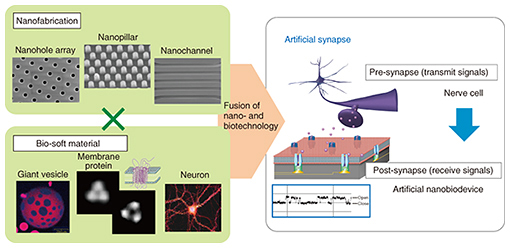
Feature Articles: Forefront Research on Bio-soft Materials Vol. 14, No. 8, pp. 7–11, Aug. 2016. https://doi.org/10.53829/ntr201608fa1 Overview of Bio-soft Material Research at NTTAbstractAt NTT Basic Research Laboratories, we are aiming to create a novel nanobiointerface that allows direct access to the human body and brain by utilizing the functions and structures of biomolecules and soft materials. We are also attempting to understand the fundamental principles of bioinformation processing by managing the combination of nanotechnology and biotechnology. The Feature Articles in this issue introduce our bio-soft material research in relation to the fabrication of nanobiodevices, which constitute one of the components of a future artificial synapse. We also present a highly sensitive and long-term stable biosensing system. Keywords: bio-soft material, nanobiointerface, bioelectrode  1. IntroductionSoft and flexible materials now play an important role in supporting modern society in the same way as hard materials such as metals, semiconductors, and ceramics. Soft materials include rubber, plastic, gel, colloid, and liquid crystal, and biological tissue also consists of large and complex soft materials. In the 20th century, hard materials played major roles in the evolution of various industries. In the 21st century, the manufacture of soft materials including their biological applications has been greatly expanded. The size, shape, and function of soft materials are easily controllable over a wide range. These materials provide us with the potential to discover unknown physical and chemical properties through the synthesis of novel soft and flexible materials or by precisely controlling their composition and structure on a nanometer-scale. In addition, these materials are not simply an interesting target for basic research; in fact, a wide range of industrial applications have also been developed. In the field of information technology and electronics, for example, plastic optical fiber and organic electro-luminescent (EL) material have been put to practical use. A tough and flexible polymer material is used as a surface coating material for mobile phones, and its surface is both highly waterproof and scratch-resistant. In recent years, it has become increasingly important to ensure that the production, use, and recycling of materials have as little effect on the environment as possible. Therefore, organic and soft materials that are people- and environment-friendly have been widely used. Another issue in the current aging society in terms of the development of medical implant materials and medical and healthcare devices is that it is important that we construct a biointerface that enables direct contact with biological systems. A sophisticated biointerface design is thus required that employs soft material with a high affinity to soft biological tissue, without causing it any damage. 2. Biointerface applicationIf we are to produce a functional biointerface that is compatible with biological systems, it is essential that we understand fundamental biostructures and design the biointerface by mimicking these biostructures. Proteins, nucleic acids, polysaccharides, and the lipid molecules of a cell membrane are often used to produce biointerfaces. These molecules have a specific feature, namely that the shape and characteristics vary greatly depending on the external environment and external stimulus. Deoxyribonucleic acid (DNA) or protein chips, various biosensors, and a drug delivery system have been developed using these molecules. In recent years, high-performance biointerfaces have been reported that can be applied to advanced medical treatments. For example, soft and highly biocompatible silicone enables us to produce an implant material that can be flexibly bent and that does not result in inflammation or rejection by the body. Damage to the spinal cord caused by an injury or an accident can result in paralysis, and to restore bodily functions, it is necessary to repair the damage site using artificial biomaterials. Thus far, it has been difficult to repair such damage. However, a research group has reported exciting results in which a paralyzed laboratory animal began to move after silicon material was embedded in the damaged part and electrical stimulation provided. Another research group has reported a functional film material that can adhere to the surface of a beating heart for a long time without falling off and without the need for sutures or adhesive. A metal or semiconductor device is printed on the film, and it features various sensor functions for detecting distortion, pH, and temperature; it also includes an actuator function that can provide electricity, heat, and a light stimulus. There is the potential to add further utilities to the functional film, and thus, it is expected that we will be able to enhance clinical applications in relation to the study and medical treatment of heart disease. Biointerface research has been conducted at NTT Basic Research Laboratories since the 1980s, and the research target is to understand neural information processing in the brain and realize a similar bioprocess using artificial devices. As an example, we have cultured a nerve cell in an artificial environment and have tried using various approaches to elucidate the nerve cell function and control neural growth. There is a synapse at the terminal of a nerve cell that transmits electrical signals to the corresponding neuron. It is well known that synapses control information processing in the brain with respect to memory and learning. The fundamental mechanism of bioelectrical signal transmission via a synapse is as follows: 1) the nerve action potential is transmitted to the synapse of a neuron; 2) the electrical signal is converted into a chemical signal (neurotransmitter), and the neurotransmitter is then released from the synapse to the outside; 3) the neurotransmitter is received via a receptor protein of the recipient cell; and finally, 4) the chemical signal is converted back to an electrical signal in the synapse of the recipient cell, and the electrical signal is then propagated in the cell. Here, the synapse that releases the neurotransmitter is called a pre-synapse, and the other part that includes a receptor protein is called a post-synapse. Information is coded by changing either the release amount or the release frequency of the neurotransmitter and modified by changing the receptor sensitivity at the synapse. Certain phenomena and functions regarding the exchange of information at the synapse are as yet unexplained. Therefore, one of our research targets involves using nanotechnology and biotechnology to artificially develop a post-synapse structure that works as an interface with a neural signal receiver. In the future, we will attempt to fabricate an artificial synapse with a simple and basic structure through the fusion of our post-synapse device and an actual nerve cell extracted from a biological pre-synapse (Fig. 1). Furthermore, we will explore the principles of bioinformation processing and clarify the information signaling mechanism in the brain. We also hope to fabricate a distinctive and valuable nanobiodevice that can work as a substitute for damaged neurotransmission elements.
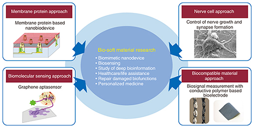
3. Bio-soft material research at NTTIn our research group, we are attempting to access fundamental bioinformation processing by utilizing a nanobiointerface or nanobiodevices, which we have fabricated by combining nanotechnology and biotechnology. We are also undertaking the research and development of novel and highly sensitive biosensors and long-term biomonitoring systems (Fig. 2). Here, we outline the research topics of each group.
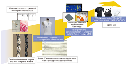
The article “Fabrication of Nanobiodevices that Utilize the Function of Membrane Proteins” [1] explains our effort to focus on a cell membrane and a membrane protein as an interface for configuring nanobiodevices. The artificially created cell membranes (artificial lipid membranes) are deposited on a silicon substrate containing a nano- or microscale hole array, and the composed structure constitutes the basic and pseudo-skeletal structure of an artificial cell. In addition, the membrane proteins, which work as a biosignal receiver, are reconstituted in the artificial lipid membrane, and this provides a platform for measuring a single protein function. The platform structure works as a post-synapse in the designed artificial synapse. The artificial lipid membrane is a soft molecular film with high fluidity, and its dynamic characteristics are an interesting research subject. In the article “Pattern Formation of Supported Lipid Bilayer for Molecular Manipulation” [2], we present an original technique for forming an artificial lipid membrane on a nanostructured surface in a precisely controlled manner. Furthermore, we describe how the lipid membrane is used as a dynamic molecule carrier. We use nerve cells to investigate the structural control of a pre-synapse that is responsible for the signal transmission of the artificial synapse. The article entitled “Neuronal Growth on Artificial Structures with Different Materials” [3] describes our study of the interfacial structure of nerve cells cultured on various substrates for which we use a technique that combines a focused ion beam with a scanning electron microscope. The topic also includes direction control in the process of nerve cell growth on a substrate with a nanopillar structure, which leads to the patterning of the nerve cell network. Moreover, in “Time-lapse Imaging of Neural Morphological Changes Relating to Cellular Functions” [4], we describe how we have used a scanning ion conductance microscope for the time-lapse imaging of the change in nerve cell structure in a physiological solution. Here, we have focused on an apoptotic process, which plays an important role in forming the most appropriate and effective neural network structure. We have clarified the correlation between morphological cell changes and the biological function by employing live imaging of the apoptotic process. In “On-chip Graphene Biosensor” [5], we describe a novel biosensor based on a graphene platform where an aptamer molecule (single-stranded DNA), which binds with a specific protein, is bound to the graphene surface. We have succeeded in selectively detecting very small amounts (below 1 μL) of proteins such as cancer or blood coagulation markers by combining a microchannel device with this biosensor. We are currently undertaking proof-of-concept experiments on the sensor, but our aim is to fabricate an on-chip biosensor that can be universally used for the qualitative and quantitative analysis of very small amounts of biomolecules. The article “Conductive Composite Material for Vital Data Measurement” [6] introduces the fabrication of a flexible and non-cytotoxic bioelectrode made of a composite consisting of silk protein and a biocompatible conductive polymer. We have used this bioelectrode to measure the electrical properties of single cells, and we have also demonstrated the long-term and stable detection of biosignals acquired from the surface of the body. 4. Evolution to wearable bioelectrodeThe bioelectrode, which is composed of two soft materials, was originally produced as a soft and fibrous microelectrode for recording very weak signals emitted by neurons in the brain. When a conventional rigid metal electrode is used, necrosis of the cells around the electrode occurs immediately. However, this soft bioelectrode has the great advantage of being hydrophilic and biologically friendly. Therefore, we can measure the nerve action potential of an experimental animal with little cell damage. By expanding this knowledge and collaborating with Toray Industries Inc., we developed a conductive fabric material called “hitoe” based on the coupling of an advanced fiber material nanofiber and a conductive polymer. “hitoe” has excellent flexibility, stretchability, breathability, and biocompatibility, and it can accurately detect biological signals such as heart rate, and provide electrocardiograms (ECG) and electromyograms (EMG) [7]. A single nanofiber is approximately 700 nm in diameter. Thanks to the fine fiber structure, adhesion with the skin is greatly improved. In addition, the high conductivity and durability of “hitoe” both help to achieve stable and long-term biological signal monitoring. Moreover, by combining “hitoe” with an undershirt, we have successfully developed a bioelectrode system that can measure the heart rate and ECG simply by having the subject wear the undershirt and by monitoring the results on a smartphone. Thanks to our collaborative work with GOLDWIN INC. and NTT DOCOMO, the undershirt has been marketed for sports applications (Fig. 3).
“hitoe” is expected to be used with a medical quality electrode for help in the early detection and treatment of heart disease. This is because “hitoe” can monitor biological information comfortably and conveniently during normal daily life without placing a burden on the wearer. Our goal is to couple “hitoe” with various wearable devices and information and communication technology, and we are therefore conducting research that will allow us to utilize bioelectrodes in a wide range of fields including medicine, healthcare, worker safety, sports, and entertainment. References
|
|||||||||||||||