 |
|||||||||||||||||||
|
|
|||||||||||||||||||
|
Special Feature: Nanobiotechnology Research Vol. 7, No. 8, pp. 25–29, Aug. 2009. https://doi.org/10.53829/ntr200908sf5 Control of Gold Nanorod Arrays Through Self-assembly of BiomoleculesAbstractThis article reports (1) the synthesis of gold nanorods coupled with biocompatible molecules and (2) the fabrication and control of gold nanorod arrays via the self-assembly of surface-anchored molecules. These successes are a major step towards various applications based on the characteristic optical properties of gold nanorods such as chemical sensors and nanophotonic, electronic, and plasmonic devices.
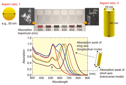
1. IntroductionThere is currently a great deal of interest in metal nanoparticles because of their shape and size-dependent optical properties, which originate from localized surface plasmon resonance (LSPR) [1]–[3]. Techniques have been developed for the synthesis and characterization of dispersed metal nanoparticles, so the next challenge is patterning and ordering these nanoscale materials into assembled structures. The controlled self-assembly of the nanoparticles on a suitable surface has many potential applications in science and technology. Furthermore, it is vital for studies on fundamental optoelectronic properties arising from collective interactions in an ordered state and for the use of nanomaterials in enhanced spectroscopy [4], (bio)chemical sensing [5], and nanophotonic, electronic, and plasmonic devices [6]. For (bio)chemical sensors, rod-like gold nanoparticles (gold nanorods: AuNRs) should provide additional benefits compared with similar-sized spherical nanoparticles because the LSPR properties of AuNRs can be continuously tuned by adjusting their aspect ratio to produce optical extinction ranging from the visible to the near-infrared (Fig. 1), where tissue transmissivity is at its highest due to the low extinction coefficients of the intrinsic chromophores. In addition, the rod-shaped geometry has inherently high sensitivity to the local dielectric environment, including the substrates, solvents, and absorbates, as well as the interparticle spacing of the AuNRs [7].
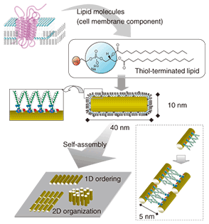
Functionalized AuNRs with specific molecular groups can provide us with precisely controlled self-assembled structures. This approach is beneficial for building blocks of AuNRs via physical and chemical affinities (covalent or noncovalent van der Waals, hydrophobic or electrostatic interactions, etc.). It is generally accepted that self-assembly with functionalized AuNRs can be an efficient means of nanofabrication owing to its simplicity, versatility, and low cost. However, previous work on AuNR self-assembly provided only ordered structures with a limited area and did not allow control of the design or interparticle distance in the AuNR architectures. 2. NanobiocompositesIn addition, despite their unique optical and nanomaterial characteristics, the application of AuNRs in the bioscience field is limited owing to the presence of a cationic detergent, which is used as a stabilizer for the AuNRs. Therefore, it is important to prepare functionalized AuNRs bearing specific biocompatible groups and to control their self-assembled structures through biomimetic, programmed intermolecular interactions. This will lead to the development of a highly sensitive biomolecular detection platform that outputs the changes in the local environment surrounding the AuNRs by utilizing their sensitive optical properties. Here, we describe the synthesis and self-assembly of two biocompatible, functionalized AuNRs coupled with 1,2-dipalmitoyl-sn-glycero-3-phosphothioethanol (thiol-phospholipid) or polyethylene glycol with a terminal thiol group (PEG-SH). We used a controlled self-assembly process to achieve precise control of the structures of the gold nanorod arrays including their dimensions, orientation, and particle gap. 3. Fabrication of gold nanorod arrays3.1 Self-assembly of complex of gold nanorod and lipidsPhospholipids are composed of a cell membrane, and their strong intermolecular interaction leads to a self-assembled lipid bilayer structure. We have successfully fabricated AuNR arrays by utilizing the self-assembly ability of surface-anchored lipids (Fig. 2).
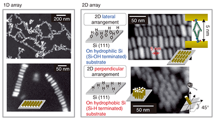
Scanning electron microscope (SEM) images of one-dimensional (1D) and two-dimensional (2D) AuNR arrays that were formed in a controllable manner when we deposited the AuNR-lipid complex on a silicon surface are shown in Fig. 3. The solvent evaporation that resulted from the spin coating used to deposit the complex led to 1D assembly with side-to-side configurations. The distance between neighboring AuNRs was uniform at around 5.0 nm, which is consistent with the thickness of the lipid bilayer. The AuNR assembly was driven by intermolecular interactions between surface-anchored lipids, and the cooperative interfacial forces that occurred during evaporation were also associated with the side-to-side ordering owing to the favorable translational entropy. Interestingly, high-density AuNR composites exhibited extensive 2D self-assembly. Furthermore, we observed anisotropically oriented AuNRs in a 2D assembly that were dependent on the nature of the Si surface (Fig. 3). On a hydrophilic Si surface (Si-OH termination), the AuNRs were organized laterally relative to the substrate. In contrast, on a hydrophobic Si surface (Si-H termination), the AuNRs were perpendicular to the substrate. In addition, the gap between neighboring AuNRs remained 5.0 nm in the 2D assembly. For the 2D array of AuNRs, our results constitute the first example of the precise control of the interparticle spacing and anisotropic orientation in a lateral or perpendicular fashion dependent on the interfacial hydrophilicity or hydrophobicity. As a control experiment, we synthesized a spherical Au nanoparticle-lipid complex and confirmed that these assembled structures formed on a hydrophilic Si substrate when we used an identical evaporation process to that used for the AuNR-lipid system. The Au nanoparticle complex was arrayed in monolayers at regular intervals of 5.1 nm and formed hexagonal structures. This indicates that our method is useful for building blocks of nanoparticles with different aspect ratios.
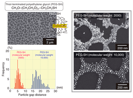
3.2 Self-assembly of complex of gold nanorod and polymersWith functionalized AuNRs with polymers, it is possible to tune the gap between adjacent AuNRs in the AuNR array. We synthesized a complex consisting of AuNR and a biocompatible polymer, polyethylene glycol with a terminal thiol group (PEG-SH), and we investigated their assembled nanostructures on silicon substrates using an SEM (Fig. 4). A self-assembled network structure was formed across all the substrates with PEG-SH-modified AuNRs. In addition, the spaces between the AuNRs were dependent on the molecular weight of the PEG-SH. When we used PEG-SH with a molecular weight of 2000, the average gap between AuNRs was estimated to be 5.52 nm in the self-assembled network structure. When we used PEG-SH with a molecular weight of 10,000, the average distance increased to 18.66 nm. If we were to use a higher-molecular-weight polymer (longer polymer chain) for the surface ligand to the AuNRs, the individual AuNR could be isolated in the polymer thin film at the single particle level. This technique is related to the tuning of the magnitude of LSPR coupling (associated with the magnitude of the local electromagnetic field enhancement) generated between AuNRs in close proximity. Therefore, we can envision the design of tailored biosensor or diagnosis biochips where we can flexibly control the effect of the surface enhancement of Raman scattering or fluorescence.
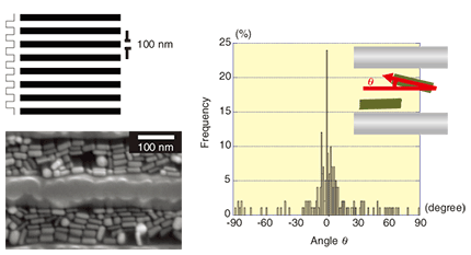
3.3 Gold nanorod arrangement using a nanopatterned silicon substrateIn terms of other AuNR arrangement methods, we attempted to use a nanopatterned silicon substrate fabricated by electron beam lithography. The AuNR-lipid complex was deposited on a 100-nm line-and- space-patterned silicon substrate, and the AuNR complex was then arranged along the trench structure of the substrate, as shown in Fig. 5. Measurement of the angle distribution of the AuNR complex, where we define the direction parallel to the trench as 0°, made it clear that almost the entire AuNR complex was located around the 0° direction. This result indicates that a nanofabricated silicon substrate can function effectively as a template for ordering an AuNR complex. We should be able to achieve the desired ordering of AuNRs with the desired orientation by using the combination of a nanoscale-controlled substrate topography and self-assembled nanoscale material. This method provides a guide to nanoscale metal wiring techniques for use on nanofabricated substrates.
4. ConclusionWe have described diverse self-assembled structures of AuNRs coupled with biocompatible molecules. AuNR-lipid complexes with anisotropically self-assembled configurations are achieved thanks to the hydrophilic or hydrophobic nature of the Si surface. The interparticle spacing of the AuNRs can be controlled by using various surface-anchored ligands with different molecular weights. The lipid- or PEG-modified AuNRs will function as anisotropic-shaped colorimetric reporters in living cells since the AuNR complex itself is biocompatible and sensitive to changes in the local environment surrounding the AuNRs. In addition, various functional biomolecules such as deoxyribonucleic acid (DNA), membrane proteins, enzymes, and antibodies can be attached to the surface of the AuNR complex and can function while retaining their physiological properties. The AuNR array on a solid surface will be utilized for surface enhanced Raman scattering or fluorescence substrates. Our future goal is to use the gold nanorod array to develop highly sensitive biochips that can detect intermolecular interactions on the single (bio)molecule scale. References
|
|||||||||||||||||||


