 |
|||||||||
|
|
|||||||||
       |
Feature Articles: Frontiers of Quantum Optics Research at NTT Basic Research Laboratories Vol. 12, No. 9, pp. 24–28, Sept. 2014. https://doi.org/10.53829/ntr201409fa5 Investigating the Properties of Glasses Using High Resolution Spectroscopy of Er3+ IonsAbstractWe have been investigating hyperfine sublevels of 167Er3+ ions doped in a silicate glass fiber with the objective of demonstrating quantum memory in the 1.5-μm telecommunication band. Memory time is determined by the stability of hyperfine sublevels (the lifetime) of 167Er3+ ions, so it is therefore necessary to clarify the lifetime properties. This article describes (i) the anomalous temperature dependence of the lifetime that was unexpectedly discovered in the lifetime measurements and (ii) the physical properties of glass that resulted in the anomalous phenomenon. Keywords: 167Er3+ ions, hyperfine sublevels, Boson peak mode
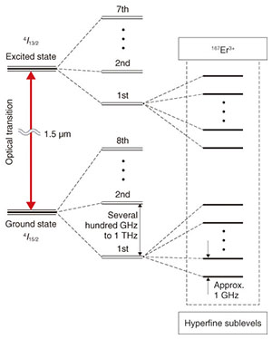
1. IntroductionThe hyperfine sublevel (hfs) properties of rare-earth (RE) ions doped in crystal have been studied intensively because RE-doped crystals are expected to be a promising material for solid-state quantum memories [1]. The memory time depends on the lifetime t1 and phase relaxation time t2 of hfs, and therefore, a systematic study was done to extend t1 and t2 to approximately seconds. NTT Basic Research Laboratories has also been studying an Er3+-doped material in order to develop a quantum memory. The hyperfine sublevels of 167Er3+ ions doped in a silicate glass fiber are one of the key properties to achieve this. There are various reasons for this, including: (i) Er3+ ions have an optical transition line (4I15/2 ↔ 4I13/2), (ii) the interaction between the Er3+ ions and light is enhanced because of the small mode field diameter (on the micrometer order), and (iii) the inhomogeneous broadening of the optical transition can provide a broadband quantum memory. We succeeded in measuring the t1 value of the hfs at 2.5–30 K using a spectroscopy method we developed, and we observed an anomalous temperature dependence whereby t1 becomes shorter as the temperature decreases from 30 K to 4 K [2]. This t1 behavior is completely different from the generally accepted view in the case of a crystalline host, where t1 becomes longer as the temperature decreases. We can consider that the glass host structure surrounding the 167Er3+ ions results in the anomalous temperature dependence. To describe this anomalous phenomenon, we examined the role of the Boson peak mode in the t1 behavior, which is a lattice vibration characteristic of glasses. 2. Er3+ ions doped in a silicate glass fiberA schematic energy-level diagram of Er3+ ions doped in a silicate glass fiber is shown in Fig. 1. The local electric field from the atoms and ions constituting the glass causes energy-level splitting to 8 levels for the ground state (4I15/2) and 7 levels for the excited state (4I13/2). The splitting energy is around several hundred GHz to 1 THz.
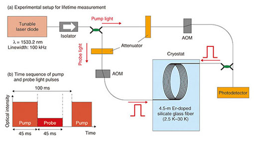
Of the many Er isotopes, only 167Er has a nuclear spin of 7/2 with an abundance ratio of 23%. The energy levels of 167Er3+ ions exhibit further splitting to 16 hyperfine sublevels with a splitting energy of around 1 GHz. This occurs because of the interaction between the electrons and the magnetic field from the nuclear spin. The optical properties of Er3+ ions doped in a silicate glass fiber have already been studied at the level of approximately 1 K [3]. Those studies revealed a homogeneous linewidth of about 10 MHz at 1 K, an inhomogeneous linewidth of about 1 THz, and an optical transition lifetime T1 of 10 ms, which is independent of temperature and equivalent to that of the case with a crystalline host. By contrast, the properties of the hfs have not yet been clarified. They exhibit large inhomogeneous broadening and may have a very short lifetime t1 compared with the crystalline host case. Consequently, it is difficult to measure these values with conventional techniques such as spectral hole burning and photon echo. 3. Measurement of lifetime t1We employed a technique that we call transient saturation spectroscopy to measure t1. Our method consists of two steps: (i) the preparation for an initial state by absorption saturation and (ii) the transition to a final state by relaxation after step (i). In step (i), we saturate the absorption of the optical transition of certain Er3+ ions that are resonant with an intense and sufficiently long pump light pulse. This process makes it possible to selectively excite particular 167Er3+ ions with an optical transition wavelength of around 1.53 µm. In a steady state, the electronic populations are distributed to the eight different hfs in the ground and excited states, as shown in Fig. 1. In step (ii), we immediately launch a weak probe light pulse with the same frequency as that of the pump light after turning off the pump light pulse. Because the intensity of the probe light pulse is sufficiently lower than that of the pump light pulse, the electronic population of each level relaxes to the ground levels with the optical transition lifetime T1 and hfs lifetime t1. Then, we can observe the transient increase in the absorption of the probe light pulse accompanied by the population relaxation, which in turn enables us to estimate T1 and t1 by measuring the decrease in the transmitted intensity of the probe light pulse. The experimental setup is shown in Fig. 2(a). We used a wavelength-tunable laser diode with its wavelength set at 1533.2 nm. The output was divided into pump and probe lights that were then pulsed by acousto-optic modulators (AOMs), where the duration and temporal position of the two pulses were controlled. The two pulses were launched into a 4.5-m Er3+-doped silicate glass fiber cooled in a cryostat system from opposite ends. The transmitted probe pulses were detected with a photodetector. The input powers of the pump and probe pulses were −6 dBm and −27 dBm, respectively. The probe pulse was launched immediately after the pump pulse had been turned off (Fig. 2(b)). The duration of each light pulse was 45 ms, and the operation period was 100 ms.
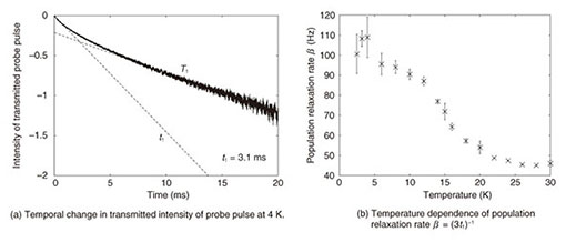
4. Lifetime properties of hyperfine sublevels of 167Er3+ ionsThe temporal change in the transmitted intensity of the probe pulse is shown in Fig. 3(a), where the temperature of the Er3+-doped silicate glass fiber was 4 K. We can clearly observe the intensity reduction with the two time constants of T1 and t1. We can estimate the t1 value at 3.1 ms at 4 K from the results. The measured population relaxation rate β (= (3t1)−1) is shown in Fig. 3(b) with respect to the temperature from 2.5 K to 30 K. The β value exhibited a nearly constant value of approximately 2π × 7.5 Hz from 30 K to 20 K. Then it increased from 2π × 8.6 Hz to 2π × 17.2 Hz with respect to the decrease in temperature from 20 K to 4 K. In the temperature region from 30 K to 4 K, we observed the anomalous temperature dependence in which the β value increased even though the temperature decreased. This observation is in direct conflict with the usual view that thermal suppression stabilizes any quantum state. In fact, with Eu3+ ions doped in the crystalline host (Eu3+:Y2SiO5), a temperature decrease from 18 K to 4 K suppresses the population relaxation of hfs from 2π × 0.1 Hz to 2π × 10−6 Hz. This comparison with the crystalline host case suggests that the anomalous temperature dependence of β can be attributed to the noncrystalline properties of a silica glass host. However, the β value decreased from 2π × 17.2 Hz to 2π × 16.0 Hz with respect to the further decrease in temperature from 4 K to 2.5 K. This behavior is in line with the usual view.
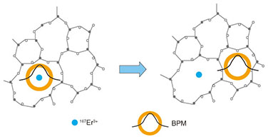
NTT Basic Research Laboratories unexpectedly found the anomalous temperature dependence of the hfs population relaxation rate β described above. We explain the physical aspect of this phenomenon in the next section. 5. Boson peak mode (BPM) in a glassTwo features that are common in glasses are an absence of translational symmetry and a disordered structure. The disordered structure results in a localized lattice vibration (phonon) mode if its wavelength is comparable to the disorder. This suggests that various low-energy vibration modes are present other than the propagation mode. In fact, it has been known for half a century that the lattice vibration in glasses shows a peak density of states (DOS) when the vibration frequency is 1 THz and the linewidth is approximately 2 THz, which is called a Boson peak. However, there is still no consensus on the origin of the lattice vibration. Several experiments have been done in the last decade using inelastic scattering of neutrons and optical Raman scattering. These experiments have gradually clarified the characteristics of the lattice vibration mode, referred to as a Boson peak mode (BPM), which may result in a Boson peak. These experiments indicated that the BPM in a silicate glass was a strongly localized mode and was relevant to the torsional motion of the SiO4 tetrahedron, which is a solid structure unit of the silicate glass. Moreover, they suggested another quite interesting feature in which the BPM that is localized at one site hops to other sites with the assistance of propagating acoustic phonons. Because the acoustic phonons increase proportionally with temperature T, the hopping probability is also proportional to T. 6. Relaxation induced by BPM in hfs of 167Er3+ ionsWe calculated the hfs population relaxation rate β for the interaction with the BPM in order to explain the obtained temperature dependence profile of β. Because the BPM in the neighbor of an Er3+ ion hops to another site at a certain time (Fig. 4), we can consider that the interaction time with the BPM is determined by the lifetime that the BPM stays at a particular site. When this lifetime increases with respect to the decrease in temperature, the long interaction with BPM promotes the relaxation of hfs of the 167Er3+ ions.
The profiles for the experimental and theoretical results are shown in Fig. 5. The profile for the theoretical results shows good agreement with that for the experimental results in the temperature region from 8 K to 30 K. This suggests that a picture of the BPM localization and hopping with the assistance of propagating phonons has been successfully obtained, which enables us to describe our experiments. The quantitative discrepancy for the temperature range of 2.5–8 K seems to be attributed to (i) underestimation of the DOS of BPM and (ii) other localized vibration modes. Nevertheless, we have succeeded in qualitatively showing the peak of the β profile.
7. Future workIn the temperature region lower than 2.5 K, the experimental and theoretical results show a significant decrease in β. We therefore intend to measure the lifetime t1 in that region and to confirm the long lifetime. After that, we will conduct quantum memory experiments. References
|
||||||||