 |
|||
|
|
|||
       |
Front-line Researchers Vol. 20, No. 10, pp. 6–11, Oct. 2022. https://doi.org/10.53829/ntr202210fr1  I Want to Reduce the Sudden Misfortune Caused by Heart Failures by Bridging the Gap between Medicine and ICTAbstractElectrocardiograms (ECGs) are widely used in medical institutions for diagnosis, vital monitoring, health checkups, and automated external defibrillators. With the development of information and communication technology (ICT) and advances in information-processing technologies such as machine learning, the application of ECGs is expanding to self-care and other fields. In today’s aging society, the need for in-home medical care and telemedicine using ECGs is rapidly increasing due to the increase in the occurrence of cardiac diseases. We interviewed Dr. Shingo Tsukada, an NTT Fellow who has been conducting interdisciplinary research that integrates medicine and ICT by drawing on his clinical experience as a physician. Keywords: tensor ECG, hitoe™, interdisciplinary research
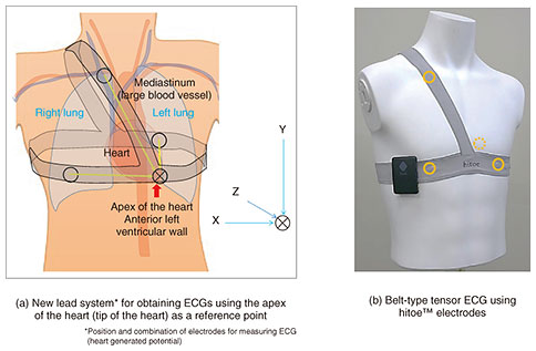
Wearable 3D ECG device with new lead system and analysis method: tensor ECG—It has been three years since our last interview. How are your research activities going? Things are going well. Using our experience in developing a wearable electrocardiogram (ECG) device using hitoe™ and by combining medical knowledge of clinical ECGs and recent information-processing technology, we are currently constructing a system for constantly measuring and analyzing ECGs. Introduced in 2014, hitoe™ is a functional fabric made by coating a conductive polymer on a cutting-edge fiber material called nanofiber fabric from Toray Industries, Inc. A clothing-type vital sensor using hitoe™ can measure biosignals with high sensitivity without burden on the wearer. In the process of our research on hitoe™, we were always conscious of ensuring our research results would be practical. The major international sporting event held in Tokyo in 2021 gave us an excellent opportunity to do just that. We have been involved in conditioning athletes taking part in swimming and cycling competitions by using the advantages of clothing-type hitoe™ capable of sensing vital data during exercise. To support two swimmers, Takeshi Kawamoto (sponsored by Toyota Motor Corporation) and Ai Soma (sponsored by Miki House), whose training base is the swimming club of Chukyo University, we proposed an exercise program for countering their decline in motor function (centered on the thorax) due to chronic muscle tension associated with their long-term and intensive training. This program promotes awareness of the functional coordination of the spine, ribs, and core muscles in a manner that restores natural and efficient body movement. In training, changes in the swimmers’ streamlined postures before and after participating in our exercise program were evaluated by photographing them from three directions and evaluating ease of breathing, lifting of the arms, and expansion and contraction of the chest circumference during breathing using a multi-sensor belt equipped with hitoe™ that measures myoelectricity, respiration, and motion. Due to the COVID-19 pandemic, we provided coaching for athlete conditioning via a smartphone-based web-conferencing system. Both Takeshi Kawamoto and Ai Soma were successful at the Japan National Championships, and I am glad that we could continue this initiative despite the pandemic. We have been involved in the measurement, analysis, and visualization of surface myoelectric potentials for cycling. Using hitoe™ as a bioelectrode, we collaborated with Bridgestone Cycle Corporation and NTT DATA to evaluate pedaling from the perspective of muscle fatigue and muscle activity in top Japanese athletes such as Eiya Hashimoto of Team Bridgestone Cycling. The athletes’ surface myoelectric potentials were measured at various locations, including the competition venue for the major international sporting event held in Tokyo, and the pedaling characteristics of each athlete were visualized using the collected measurement data. The visualized characteristics were then fed back to the athletes for discussions based on their intuitions and subjective opinions in a manner that enabled us to identify points for strengthening. Knowing the status of muscle activity during competition is an important factor in improving and conditioning an athlete’s performance. The surface myoelectric potentials measured using hitoe™ can monitor muscle activity from the surface of the body, thus are considered effective for on-site analysis of exercise because they can be measured by having the user simply change clothes in a manner that imposes little burden on the body. Through the above-described initiatives for cycling, we confirmed the usefulness of hitoe™ as a bioelectrode. —New research themes derived from hitoe™ have been created, right? Having previously worked as a physician, I joined NTT mid-career in 2010 and started my research in a new field almost from scratch. I was in charge of everything from the invention of bioelectrodes to the creation of fabrics for bioelectrodes that enable constant monitoring of heartbeats and ECGs just by wearing it and implementation of these technologies. Although we have proposed various bioelectrodes and wearable devices, only a small percentage have been implemented, and other companies are in the same situation. Fortunately, my proposal was a new type of a wearable biometric sensor, so it attracted attention in Japan and from abroad, and I was busy flying all over the world to promote it. In the midst of the stress brought on by that travelling, and because doctors often neglect their own health, I suddenly suffered a heart attack. I was shocked by the ECG obtained using the wearable ECG device that I was studying at the time and by the ECG obtained during the provocation test conducted for precise examination at the hospital. Compared with the intense subjective symptoms I experienced during the heart attack, the abnormality (signal waveform distortion) shown by the ECGs were extremely small. Regardless of the fact that wearable ECG devices have been studied with the aim of avoiding heart attacks and enabling early diagnosis of heart failure, conventional methods of interpreting an ECG (which are the criteria for determining normal or abnormal ECG) have their limitations. I became aware of the existence of small abnormalities that could be overlooked and cause heart attacks even when using these wearable ECG devices. Overcoming this limitation will require innovative techniques for ECG analysis by using information-processing technology. The behavioral restrictions placed on people due to the COVID-19 pandemic allowed me to change my busy life and take time to focus on this issue and create tensor ECG, a new method of analyzing ECGs. The experience of a clinician and non-specialist in information processing and statistics was useful—Your unfortunate experience was behind the development of a new method for ECG analysis. Could you tell us more about tensor ECG? An ECG is a time-varying representation of potential differences acquired from multiple bioelectrodes placed on the chest, extremities, and other parts of the body. The chest electrodes are less affected by body movement, thus produce relatively large cardiac potentials. However, the other electrodes are significantly affected by body movements; accordingly, ECG measurements are basically conducted with the user in a resting state. To find minute abnormalities in an ECG (i.e., distortion of the waveform), it is necessary to stably record the ECG over a long period. We therefore devised a wearable ECG device with a new lead system, i.e., placing electrodes in three linearly independent directions using the apex region of the heart (i.e., from the apex to anterior left ventricular wall), where the heart is closest to the rib cage, as a reference point (Fig. 1). Since the heart and its movement are three-dimensional (3D), a 3D ECG corresponding to the movement of the heart is obtained from electrodes placed three-dimensionally around the heart. To improve wearability of the electrodes and minimize the effects of body movement on the position of the electrodes, the electrodes and wiring are integrated in an elastic belt, which can be easily fitted to the user’s body by simply tightening the shoulder and waist belts. We also developed a polygraph (mechanocardiogram) that simultaneously measures cardiac output and deep-vascular pulse waves.
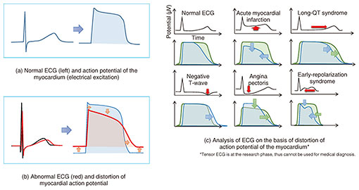
Conventional criteria for normal ECG include a wide range of values for waveforms and intervals, and individual criteria are currently used for each disease or pattern. Even when a person has a heart attack (angina pectoris or other abnormality), their waveform may only be slightly distorted. A method for quantitatively evaluating abnormalities in atypical ECGs has not been developed, and in some cases, abnormalities have been judged to be normal. An ECG is a recording made from the body surface of electrical potentials generated by the excitation (called action potentials) of numerous myocardial cells. The action potential of myocardial cells cannot be measured from the body surface. Therefore, we devised a method of (i) statistically modeling the timing of changes in the action potential by using a Gaussian distribution and (ii) estimating the collective transition of action potentials of the myocardial cells (cardiac muscle) from an ECG. This method makes it possible to resolve, amplify, and visualize atypical distortions in an ECG (Fig. 2).
We are currently verifying whether the parameters obtained with tensor ECG are effective indicators for classifying complex cardiac abnormalities and quantifying and evaluating minute distortions in ECGs that have been hitherto overlooked in a unified manner. We hope that this method will be useful for diagnosing arrhythmias (irregular heartbeat) associated with heart failure, ischemic heart disease, and sudden cardiac death. —Has this type of analysis of ECGs been done before? I have checked previous papers and prior patents regarding tensor ECG, but I found no similar cases, so I believe the novelty of this development is high. In Europe and the US, where heart failure and heart attacks are the leading causes of death, research in this field is thriving, and competition in research and development has been intensifying as Apple and other companies enter the field. Many of these research efforts use machine learning and artificial intelligence (AI) to automatically determine abnormal ECGs and attempt to detect abnormalities that could not be detected before. However, ECG interpretation using AI faces certain issues, for example, the dependence of the ECG data used to train the AI, ambiguity of causal relationships between ECG data and judgement, and necessity of preparing a large amount of ECG data. The so-called inverse problem in electrocardiography, which involves calculating myocardial action potentials from ECGs and analyzing abnormalities, has been regarded as a difficult problem with no solution. Moreover, abnormalities in ECGs often involve only slight distortions of shape or changes in potential. However, I know that certain clinicians are good at reading subtle changes in ECGs. I hypothesized that they identify abnormalities on the basis of their experience of examining ECGs of many patients. To solve the above problem, I studied information processing and statistics. The medical science that I studied at university was a far cry from today’s information processing, so I had to start from scratch again. However, one day, while relearning about the Gaussian distribution in statistics, I had an inspiration of a very simple model equation and found that it could solve the problem, albeit with some limitations. Perhaps it was my amateur ideas concerning information processing and statistics that worked. The researcher’s mission is to find and nurture new seeds for a new era—In our last interview, you mentioned that your dream is to develop advanced medical devices for detecting signs of illness, and your dream looks as if it will be coming true very soon. I do not currently know if tensor ECG is the correct method or useful for clinical medicine. If this method is validated, it would truly be a dream come true as well as a major breakthrough. We are now entering the stress-test phase, and we face a number of technical challenges and must overcome a number of major hurdles before we can start implementing it. We have just started clinical research with tensor ECG, but if its effectiveness and value are recognized, it could be adopted as a method for analyzing ECGs in about three to five years. However, AI diagnosis of ECGs is developing at a breakneck pace, so it is possible that tensor ECG will lose out. Tensor ECG is a versatile analysis method applicable to all types of ECGs. If its clinical efficacy is confirmed, it could contribute to society by enabling precise analysis of a person’s ECGs and detection of signs of heart disease by simply clicking on a website in, say, ten years or so. Academically speaking, I’m considering submitting papers to interdisciplinary journals that cover topics between information processing and medicine. I realize that many of today’s research problems cannot be solved by research in a single field of expertise. In fact, in Europe and the US, cross-disciplines or double majors are common—that is, researchers and specialists who have mastered one field of expertise pursue another field of expertise—or work closely with those with another field of expertise to solve problems. However, I feel that in Japan, cross-disciplinary and interdisciplinary studies have not progressed, and such researchers of my generation are few and far between. My work as a researcher at NTT spans the fields of medicine, physiology, medical engineering, and bioinstrumentation and information processing. I hope to help build a bridge among experts in different fields and address social issues such as rising medical costs. —What do you value as a researcher? I believe that the role of a researcher is to find new seeds and nurture them for the next generation. In every field, there are research themes that attract attention at certain times, but not all are necessarily connected to important issues. Therefore, I want to approach my research with an eye to the importance of the theme and the social necessity of research on that theme. While it is essential to enhance one’s expertise, I also want to actively engage in interdisciplinary exchanges and hands-on experience. Through various exchanges, I can understand the difficulties and technical bottlenecks in each field, and unique ideas can be born from them. To facilitate such interaction and communication, I hope to continue to be a “handyman” who is easy to talk to, as I said in our last interview. From my experience as a physician dealing with patients, I believe that happiness is not simply brought by success, achievement, status, and so on; rather, by being blessed with family, friends, and colleagues and interacting with others. This feeling is one that I share with many of the health-care professionals I have worked with. It seems that researchers who have good relationships with those around them are more likely to be able to continue their research activities and achieve more consistent results. Of course, some researchers have achieved remarkable results in their fields of expertise on their own. Research styles can vary individually, but I’d like to continue my research activities while engaging with a variety of people and helping one another. Researchers are evaluated on the basis of the number of papers they have published, the impact factor of those publications, and other indicators, and I feel that what researchers who have achieved outstanding results have in common is being good communicators. That means they have outstanding communication skills and are well versed in fields outside their expertise; for example, they can relate to topics and difficulties in other fields. I hope to be the same way; that is, I want to continue to conduct research that will lead to a brighter future in a manner that enables my research results to be as much use to society as possible and allows young researchers to have more opportunities to be active. ■Interviewee profileShingo Tsukada graduated from Toyama University School of Medicine and received a medical license in 1990. He also received a Ph.D. in medicine from the University of Tsukuba in 2003. He was a visiting researcher at the University of California San Diego from 2003 to 2005. He joined NTT Basic Research Laboratories in 2010 as a research specialist. He has been studying cardio vascular function and neuronal regulation. His current interests include the detection of biomedical signals and functional modification using novel wearable-type and implant-type bioelectrodes based on the composites of conductive polymers with various fibers and textiles. He is an inventor of the textile bioelectrode hitoe™. He is a member of the Physiological Society of Japan, the Japan Society of Applied Physics, the Japanese Circulation Society, the Japanese Orthopedic Association, and the Japanese Association of Rehabilitation Medicine. |
||